dentigerous cyst Nature, diagnosis and treatment
dentigerous cyst (DC)
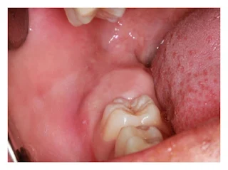
A dentigerous cyst (DC) is one that is formed by follicle expansion of an unerupted tooth enclosing its crown.
Tooth follicle (tooth sac) normally found surrounding developing tooth bud normaly 2-3mm on radiograph , but abnormal expantion may leads to DC formation .
Dentigerous cysts are the second most common type of odontogenic cyst, which is a fluid-filled sac that develops in the jaw bone and soft tissue. They form over the top of an unerupted tooth, or partially erupted tooth, usually one of your molars or canines. While dentigerous cysts are benign, they can lead to complications, such as infection, if left untreated.
DCs Accounting for 25% of all the cysts , making it one of the frequently occurring developmental cysts of jaw.
Although noticed in wide age range , most commonly seen in between 20 to 30 years of life, less frequently below 10 years of age. Majority of the cases shows association with the impacted or unerupted mandibular molars,
second most common site is maxillary canines followed by maxillary molars. Most of the DCs are painless unless secondarily infected, however mainly noticed during routine radiographic examination. Larger DCs sometimes can resemble aggressive lesions like keratocystic odontogenic tumor and ameloblastoma. Entities like unicystic ameloblastoma (50% of the cases) and ameloblastic fibroma(75% of the cases) have similarities in some aspects to DC such as, predilection to occur in children and young adolescents and frequently seen in association with unerupted or impacted tooth (75% of ameloblastic fibromas).
A careful evaluation of clinical, radiological and histopathological differential diagnosis is needed before scheduling the surgery. The timely diagnosis and treatment should be done as untreated cases of dentigerous cyst can lead to complication such as, bone deformation, loss of permanent tooth and may also develop into odontogenic tumors and carcinomas.
INVESTIGATIONS AND DIFFERENTIAL DIAGNOSIS
Clinical differential diagnosis included periapical cyst
keratocystic odontogenic cyst (KOT)
and ameloblastoma.
How is it diagnosed?
Small dentigerous cysts often go unnoticed until you have a dental X-ray. If your dentist notices an unusual spot on your dental X-ray, they may use a CT scan or MRI scan to make sure it’s not another type of cyst, such as a periapical cyst or an aneurysmal bone cyst.
In some cases, including when the cyst is larger, your dentist may be able to diagnose a dentigerous cyst just by looking at it.
Root resorption is seen with respect to the neiboring tooth .
The most favourable radiographic differential diagnosis for DC, ameloblastic fibroma , unicystic ameloblastoma, KOT, and adenomatoid odontogenic tumor (AOT)
DC was the first choice of diagnosis as the radiograph revealed unilocular radiolucency surrounding the neck of the crown of an unerupted tooth, with diffuse and thin corticated borders, which are the radiographical features usually seen in DC. DC has to be differentiated from the hyperplastic follicle. If the follicular space is above 5 mm DC can suspected , as the normal follicular space is 2-3mm .
But other entities like ameloblasctic fibroma, unicystic ameloblastoma, KOT and AOT has to be ruled out before considering the treatment options.
Ameloblastic fibroma is one of the differential diagnosis for this case, as OPG showed unilocular radiolucency surrounding the neck of the tooth . Usually ameloblastic fibroma appears as a unilocular radiolucency and sometimes multilocular. It usually shows well demarcated sclerotic border and sometime associated with unerupted or displaced tooth. We had to wait for the histopathological findings to rule out ameloblastic fibroma.
Unicystic ameloblastoma (UA) of dentigerous variant shows unilocular radiolucency in association with an impacted tooth. The involved tooth mainly would be mandibular third molar followed by mandibular canines in case of unicystic ameloblastoma. Knife edge root resorption and destruction of the anterior border of the ramus are also the characteristic features of the UA.
Unicystic ameloblastoma is also one of the probable and important radiographical differential diagnosis .To rule out this entity histopathology report is needed.
KOT radiographically seen as unilocular or multilocular radiolucency surrounded by corticated margins with slight tendency towards unilocular radiolucency. Where 30% of the cases are associated with the unerupted teeth, mainly third molars. The DC of bigger size always pose problem in diagnosis between KOT and DC. One preculiar feature of KOT is antero-posterior expansion with considerable mesiodistal extension.
Radiographic findings of AOT sometimes resemble dentigerous cyst. Follicular type of AOT which is seen in association with the unerupted tooth could be the potential differential diagnostic entity . AOT is seen as clinically, periapical cyst was ruled out.
Orthopantamograph (OPG) is advised. The OPG findings showed, a well-defined unilocular radiolucent area in association with the crown of an unerupted tooth with diffuse corticated border. On the unilocular radiolucency or sometimes mixed radiopaque - radiolucent lesion with well defined sclerotic or corticated border.
How is it treated?
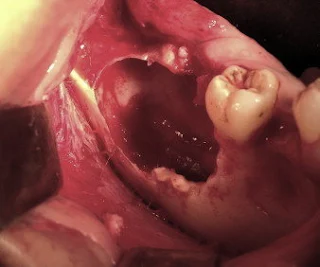
Treating a dentigerous cyst depends on its size. If it’s small, your dentist might be able to surgically remove it along with the affected tooth by enucleation. In other cases, they might use a technique called marsupialization.
Marsupialization involves cutting open the cyst so it can drain. Once the fluid has drained, stitches are added to the edges of the incision to keep it open, which prevents another cyst from growing there.


This is an interesting publication about the nature of dentigerous cyst, diagnosis and treatment. There is adequate explanation provided within the writing, thoroughly explaining the process of diagnosing such a condition and the steps taken to treat and restore normal gum function. Keep providing updates to your blog. Have a wonderful rest of the day.
ReplyDeleteDentist Philadelphia