Cleaning and Shaping principles ,basic definitions and irrigations used
Cleaning and Shaping principles , basic definitions and irrigations used
Successful root canal treatment is based on establishing an accurate diagnosis and developing an appropriate treatment plan; applying knowledge of tooth anatomy and morphology (shape); and performing the debridement, disinfection, and obturation of the entire root canal system. Initially emphasis was on obturation and sealing the radicular space. Adequate cleaning and shaping and establishing a coronal seal are the essential elements for successful treatment with obturation being less important for short term success. Sealing the canal space following cleaning and shaping will entomb any remaining organisms and, with the coronal seal, prevent re-contamination of the canal and periradicular tissues.
principles of cleaning
Success of root canal treatment in a tooth with a vital pulp is higher than that of a tooth that is necrotic with periradicular pathosis. The difference is the persistent irritation of necrotic tissue remnants, and the inability to remove the microorganisms and their by-products. The most significant factors affecting this process are tooth anatomy and morphology, and the instruments and irrigants available for treatment. Instruments must contact and plane the canal walls to debride the canal. Morphologic factors such as lateral and accessory canals, canal curvatures, canal wall irregularities, fins, cul-de-sacs and ishmuses make total debridement virtually impossible. Therefore the goal of cleaning not total elimination of the irritants but it is to reduce the irritants.
 |
| examples of canal irregularities |
Currently there are no reliable methods to assess cleaning. The presence of clean dentinal shavings, the color of the irrigant, and canal enlargement three file sizes beyond the first instrument to bind have been used to assess the adequacy; however, these do not correlate well with debridement. Obtaining glassy smooth walls is a preferred indicator.
Principles of Shaping
The purpose of shaping is to
1) facilitate cleaning and
2) provide space for placing the obturating materials.
The main objective of shaping is to (maintain or develop a continuously tapering funnel from the canal orifice to the apex).
This decreases procedural errors when cleaning and enlarging apically. The degree of enlargement is often dictated by the method of obturation. For lateral compaction of gutta percha the canal should be enlarged sufficiently to permit placement of the spreader to within 1-2 millimeters of the corrected working length. There is a correlation between the depth of spreader penetration and the apical seal. For warm vertical compaction techniques the coronal enlargement must permit the placement of the pluggers to within 3 to 5 mm of the corrected working length.
The degree of shaping is determined by the preoperative root dimension, the obturation technique, and the restorative treatment plan. Narrow thin roots such as the mandibular incisors cannot be enlarged to the same degree as more bulky roots such as the maxillary central incisors. Post placement is also a determining factor in the amount of coronal dentin removal.
Apical Patency
small file 10 or 15 used to and beyond the apex frequently
Apical patency has been advocated during cleaning and shaping procedures 1) to ensure working length is not lost and that 2) the apical portion of the root is not packed with tissue, dentin debris and bacteria . Concerns regarding extrusion of dentinal debris, bacteria and irrigants have been raised. Seeding the periradicular tissues with microorganisms may occur.
The apical patency concept also has been advocated to 3) facilitate apical preparation. Extending the file beyond the apex increases the diameter of the canal at working length consistent with the instrument taper. The value of maintaining patency to prevent transportation is questionable and it does not result in bacterial reduction when compared to not maintaining patency. Small files are not effective in debridement .
Ultrasonics
Ultrasonics are used for
-cleaning and shaping,
-removal of materials from the canal,
-removal of posts and silver cones,
-thermoplastic obturation, and
- root end preparation during surgery.
The main advantage to cleaning and shaping with ultrasonics is acoustic micro streaming. This is described as a complex steady-state streaming patterns in a vortex like motion or eddy flows formed close to the instrument. Agitation of the irrigant with an ultrasonically activated file after completion of cleaning and shaping has the benefit of increasing the effectiveness of the solution.
Initially it was proposed that ultrasonics could clean the canal without procedural errors such as apical transportation and remove the smear layer. However later studies failed to confirm these results.
IRRIGANTS AND LUBRICANTS
Irrigation does not completely debride the canal. Sodium hypochlorite will not remove tissue from areas that are not touched by files . In fact no techniques appear able to completely clean the root canal space. Frequent irrigation is necessary to flush and remove the debris generated by the mechanical action of the instruments.
Properties of an ideal irrigant
Organic tissue solvent
Inorganic tissue solvent
Antimicrobial action
Non-toxic
Low Surface Tension
Lubricant
Antimicrobial action
Sodium Hypochlorite
The most common irrigant is sodium hypochlorite (household bleach). Advantages to sodium hypochlorite include
the mechanical flushing of debris from the canal
the ability of the solution to dissolve vital and necrotic tissue,
the antimicrobial action of the solution
the lubricating action . In addition it is inexpensive and readily available.
Free chlorine in sodium hypochlorite dissolves necrotic tissue by breaking down proteins into amino acids. There is no proven appropriate concentration of sodium hypochlorite, but concentrations ranging form 0.5% to 5.25% have been recommended. A common concentration is 2.5%; which decreases the potential for toxicity while still maintaining some tissue dissolving and antimicrobial activity. Since the action of the irrigant is related to the amount of free chlorine, decreasing the concentration can be compensated by increasing the volume. Warming the solution can also increase effectiveness of the solution.
Because of toxicity, extrusion is to be avoided.The irrigating needle must be placed loosely in the canal .
Insertion to binding and slight withdrawal minimizes the potential for possible extrusion and a “sodium hypochlorite accident” . Special care should be exercised when irrigating a canal with an open apex. To control the depth of insertion the needle is bent slightly at the appropriate length or a rubber stopper placed on the needle.
The irrigant does not move apically more than one millimeter beyond the irrigation tip so deep placement with small gauge needles enhances irrigation Unfortunately the small bore can easily clog, so aspiration after each use is recommended. During rinsing, the needle is moved up and down constantly to produce agitation and prevent binding or wedging of the needle.
Chlorhexidine
Chlorhexidine possesses a broad spectrum of antimicrobial activity, provides a sustained action and has little toxicity. Two percent chlorhexadine has similar antimicrobial action as 5.25% sodium hypochlorite and is more effective against enterococcus faecalis. Sodium hypochlorite and chlorhexadine are synergistic in their ability to eliminate microorganisms. A disadvantage of chlorhexadine is its inability to dissolve necrotic tissue and remove the smear layer.
LUBRICANTS
Lubricants facilitate file manipulation during cleaning and shaping. They are an aid in initial canal negotiation especially in small constricted canals without taper. They reduce torsional forces on the instruments and decrease the potential for fracture.
Glycerin is a mild alcohol that is inexpensive, nontoxic, aseptic, and somewhat soluble. A small amount can be placed along the shaft of the file or deposited in the canal orifice. Counterclockwise rotation of the file carries the material apically. The file can then be worked to place using a watch winding or “twiddling” motion.
Paste lubricants can incorporate chelators. One advantage to paste lubricants is that they can suspend dentinal debris and prevent apical compaction. One proprietary product consists of glycol, urea peroxide and ethylenediaminetetraacetic acid (EDTA) in a special water soluble base. It has been demonstrated to exhibit an antimicrobial action. Another type is composed of 19% EDTA in a water soluble viscous solution.
A disadvantage to these EDTA compounds appears to be the deactivation of sodium hypochlorite by reducing the available chlorine and potential toxicity. The addition of EDTA to the lubricants has not proven to be effective. In general files remove dentin faster than the chelators can soften the canal walls. Aqueous solutions such as sodium hypochlorite should be used instead of paste lubricants when using nickel-titanium rotary techniques to reduce torque.
EDTA
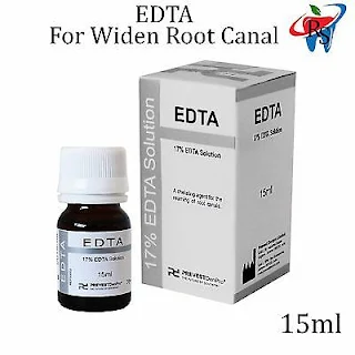
Removal of the smear layer is accomplished with acids or other chelating agents such as ethylenediamine tetracetic acid (EDTA) following cleaning and shaping. Irrigation with 17% EDTA for one minute followed by a final rinse with sodium hypochlorite is a recommended method. Chelators remove the inorganic components leaving the organic tissue elements intact. Sodium hypochlorite is then necessary for removal of the remaining organic components. Citric acid has also been shown to be an effective method for removing the smear layer as has tetracycline.
Demineralization results in removal of the smear layer and plugs, and enlargement of the tubules. The action is most effective in the coronal and middle thirds of the canal and reduced apically. Reduced activity may be a reflection of canal size or anatomical variations such as irregular or sclerotic tubules. The variable structure of the apical region presents a challenge during endodontic obturation with adhesive materials.
The recommended time for removal of the smear layer with EDTA is 1 minute. The small particles of the smear layer are primarily inorganic with a high surface to mass ratio which facilitates removal by acids and chelators. EDTA exposure over 10 minutes causes excessive removal of both peritubular and intratubular dentin.
SMEAR LAYER
During the cleaning and shaping, organic pulpal materials and inorganic dentinal debris accummulates on the radicular canal wall producing a an amorphous irregular smear layer With pulp necrosis, the smear layer may be contaminated with bacteria and their metabolic by-products. The smear layer is superficial with a thickness of 1-5 microns and debris can be packed into the dentinal tubules varying distances.
There does not appear to be a consensus on removing the smear layer prior to obturation. The advantages and disadvantages of the smear layer removal remain controversial; however, evidence supports removing the smear layer prior to obturation.The organic debris present in the smear layer might constitute substrate for bacterial growth and it has been suggested that the smear layer prohibits sealer contact with the canal wall and permits leakage. In addition, viable microorganisms in the dentinal tubules may use the smear layer as a substrate for sustained growth. When the smear layer is not removed, it may slowly disintegrate with leaking obturation materials, or it may be removed by acids and enzymes that are produced by viable bacteria left in the tubules or enter via coronal leakage. The presence of a smear layer may also interfere with the action and effectiveness of root canal irrigants and inter-appointment disinfectants.
MTAD
An alternative method for removing the smear layer employs the use of a mixture of a tetracycline isomer, an acid, and a detergent (MTAD) as a final rise to remove the smear layer. The effectiveness of MTAD to completely remove the smear layer is enhanced when low concentrations of NaOCl are used as an intracanal irrigant before the use of MTAD. A 1.3% concentration is recommended. MTAD may be superior to sodium hypochlorite in antimicrobial action. MTAD has been shown to be effective in killing E. faecalis, an organism commonly found in failing cases, and may prove beneficial during retreatment. It is biocompatible, does not alter the physical properties of the dentin and it enhances bond strength.
Related topics


Comments